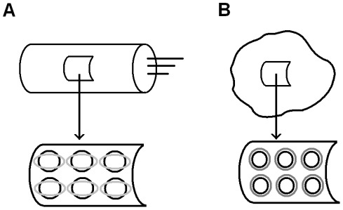
Figure 1. Cellular membranes containing the protein prestin.
A) Upper panel: outer hair cell. Lower panel: representative cut from the outer hair cell composite membrane (wall). The molecules of prestin distributed along the membrane (cell) surface undergo conformational changes schematically sketched as black circles transforming into gray ellipses. This elliptical shape is consistent with the active strain being compressive in the circumferential direction and extensive in the longitudinal direction along the cylindrical cell whose wall has anisotropic properties [37]. B) Upper panel: cell transfected with prestin and representative section of its membrane. Lower panel: representative section of its membrane. The molecules of prestin distributed along the membrane (cell) surface undergo conformational changes schematically sketched as black circles transforming into gray circles.

