Figure 6.
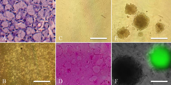
Adult mouse lacrimal gland epithelial cell cultures with CT. H&E staining of the adult lacrimal gland showing regular acinar unit structures (A). Primary cultures of adult lacrimal gland epithelial cells after 10 days showing a cobblestone structure (B). After subculture, cells at passage 1 showed similar cell morphology (C). Clear colony formation was generated on 3T3 feeder layers after 12 days (D). Spheres were also generated from adult lacrimal gland cells after 10 days (E), and were generated from GFP positive or GFP negative cells (F). Scale bars, (B, C, E) 100 µm; (F) 50 µm.

