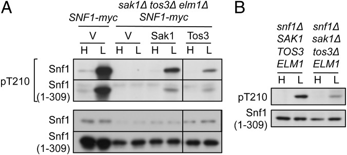
Fig. 4.
Phosphorylation of Snf1(1–309) by Snf1-activating kinases. Preparation of cells and immunoblot analysis were as in Fig. 1. (A) SNF1-myc cells and sak1Δ tos3Δ elm1Δ SNF1-myc cells expressed Snf1(1–309)-myc and Sak1-5xV5 or Tos3-5xV5 from the native promoters on centromeric plasmids pRS316-Sak1-5V5 (56) or pYL388 (43), respectively, or carried vector (V). All lanes are from the same blot. (B) snf1Δ sak1Δ tos3Δ ELM1 cells and control snf1Δ cells expressed Snf1(1–309)-myc. Control cells carrying vector showed no phosphorylation.

