Fig. 3.
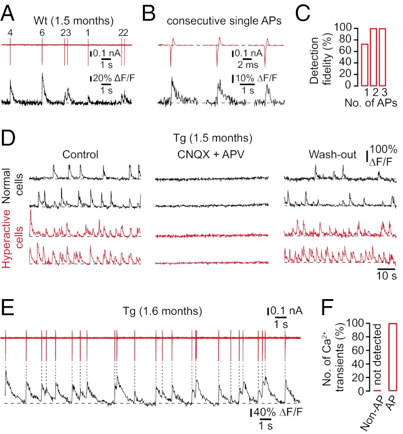
Cellular mechanisms of spontaneous Ca2+ transients in wild-type and transgenic mice. (A) Simultaneous in vivo recordings of spontaneous Ca2+ transients (black trace) and underlying action potential (AP) firing (red trace; number of APs indicated) in a cell-attached configuration from a CA1 neuron in a wild-type mouse. (B) Examples of spontaneous Ca2+ transients (black trace) evoked from three consecutive single APs (red trace) in a WT mouse. (C) Fractions of single APs and trains of APs optically detected in CA1 neurons of WT mice (n = 15 cells in three mice). (D) Spontaneous Ca2+ transients in normal (black traces) and hyperactive (red traces) CA1 neurons of a transgenic mouse before, during, and after local application of CNQX and APV. (E) Simultaneous in vivo recordings of spontaneous Ca2+ transients (black trace) and underlying action potential firing (red trace) in a cell-attached configuration from a CA1 neuron in a tg mouse. (F) Number of spontaneous Ca2+ transients triggered (AP) and not triggered (non-AP) by action potential firing in tg mice (n = 13 cells in three mice).

