Figure 1.
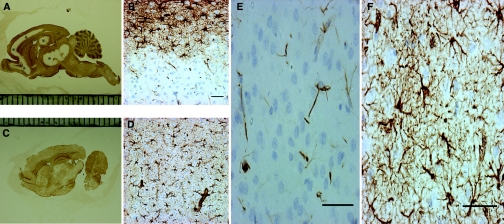
Rapid correction of chronic hyponatremia induces glial fibrillary acidic protein (GFAP) loss. In (A), immunohistochemistry for GFAP in rats 2 days after hyponatremia correction reveals a salient lack of immunoreactivity in the cortical regions, hippocampus, and basal ganglia (marked with *). At a higher magnification, in (B) the cortex of corrected rats shows a crisp demarcation between the area of GFAP reactivity and the region negative for GFAP. This contrasts with the normal GFAP (C and D) staining that was observed in uncorrected rats. In corrected rats, regions lacking macroscopic GFAP staining did not display any GFAP-positive cells (E) and only a limited amount of immunoreactive cellular debris. In contrast, as seen in (F), uncorrected control rats show a homogenous, star-shaped cellular GFAP staining pattern. The scale bar is 50 μm.

