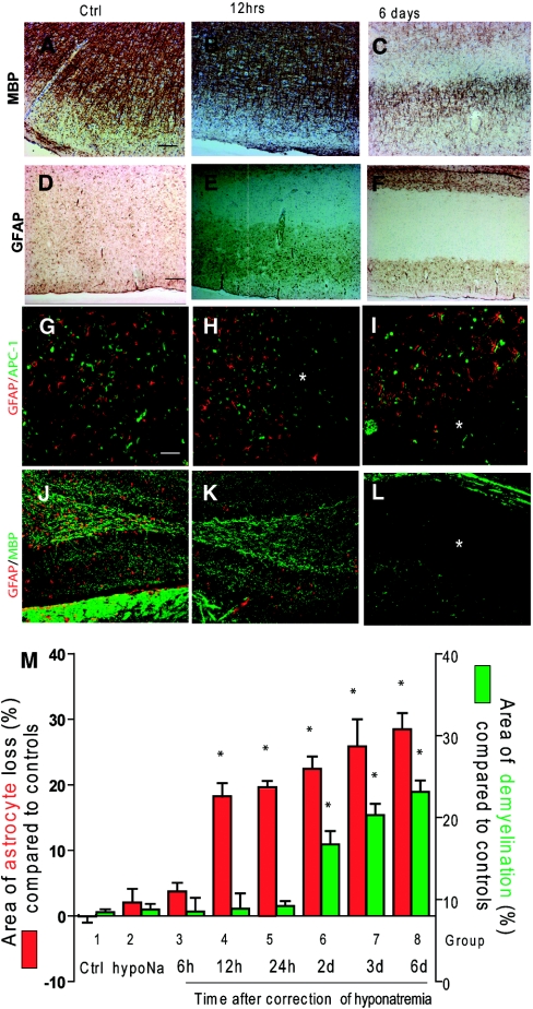
Figure 3.
Loss of astrocytes occurs before demyelination after excessive correction of chronic hyponatremia. Myelin labeling (anti-MBP) in the cortex of uncorrected hyponatremic rats (A) and 12 hours (B) postcorrection did not show any reduction of MBP staining in uncorrected rats or 12 hours after hyponatremia correction. However, at 6 days postcorrection (C), myelin staining was reduced in the subcortical regions. Homogenous GFAP staining was found in uncorrected controls (D), but there was a marked loss of subcortical GFAP staining as early as 12 hours after the correction (E) that persisted for up to 6 days postcorrection (F). Double immunofluorescence detection of astrocytes (GFAP in red) and oligodendrocytes (adenomatous polyposis coli (APC) clone CC1 in green) shows a normal density of astrocytes and oligodendrocytes in uncorrected control rats (G). Astrocyte loss occurred 12 hours after the correction of hyponatremia (*) without evident oligodendrocyte death (H), whereas both astrocytes and oligodendrocytes were lost 6 days postcorrection, which is shown in the lower half of the image (*) (I). In (J) and (K), double immunofluorescence showing the preservation of myelin (MBP in red) in the hippocampus at 12 hours postcorrection despite astrocyte death (GFAP in green). Six days after correction (L), myelin loss had occurred in the astrocyte-depleted regions (*). The scale bar is 50 μm. In the graph (M), astrocyte loss (GFAP-negative brain surface area in green) and demyelination (MBP-negative brain surface area in red) were quantified after hyponatremia correction by computer-assisted image analysis, showing the progression of astrocyte and myelin damage over the 6 days after correction of serum sodium. Each group represents a different time point postcorrection. (*, P < 0.001 compared with controls, ANOVA test, n = 6 for each group). Ctrl, control; GFAP, glial fibrillary acidic protein; MBP, myelin basic protein; hypoNa, hyponatremia.

