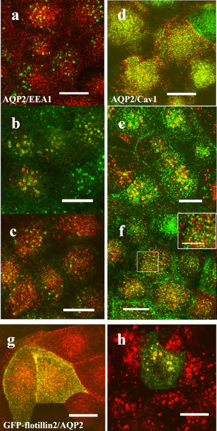
Fig. 6.
AQP2 is internalized to the same compartment with caveolin-1 and flotillin-2. (a–f) MDCK-hAQP2 cells were seeded on coverslips and subjected to forskolin treatment for 30 min (a, d) and subsequent washing and incubation without forskolin for 30 min (b, e) or for 2 hr (c, f). Double-immunofluorescence labeling for AQP2 and EEA1 (a–c) or for AQP2 and caveolin-1 (d–f) was carried out and their localization was observed with a laser confocal microscope. Projection images of 4 consecutive confocal images (0.4 µm intervals) are shown. AQP2 is shown in red. EEA1 and caveolin-1 are shown in green. An enlarged view of the rectangle area is shown in the inset (f). Both AQP2 and caveolin-1 are internalized in the EEA1-positive compartment 30 min after washing and then differentially localized at 2 hr. g, h: MDCK-hAQP2 cells transiently transfected with EGFP-flotillin-2 were treated with forskolin for 30 min (g) and then washed and incubated without forskolin for 30 min (h). Localization of AQP2 and flotillin-2 was observed with a laser confocal microscope. Projection images of 4 consecutive confocal images (0.4 µm intervals) are shown. AQP2 is shown in red. EGFP fluorescence is shown in green. Both AQP2 and EGFP-flotillin-2 are internalized in the same compartment. Bars=10 µm (5 µm in inset).

