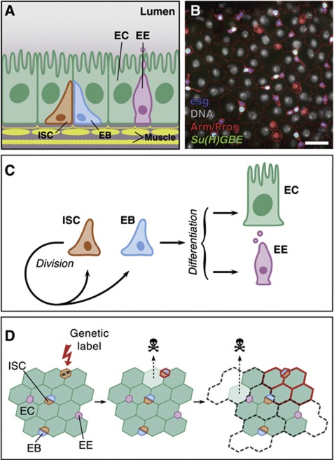
Figure 1.
Composition and homeostasis of the adult posterior midgut. (A) Organization of the midgut epithelium in enterocytes (EC), enteroendocrine cells (EE), enteroblasts (EB) and intestinal stem cells (ISC), wrapped in muscle fibres. (B) View of the Drosophila midgut epithelium showing expression of Arm and Pros (red), esg-lacZ (blue), Su(H)GBE-GFP:nls (green) and DNA (grey pseudocolour). Separate channels are in Supplementary Figure S1. Bar: 20 μm. (C) Model of midgut homeostasis based on the asymmetric fate outcome of ISC division, and EB acting as a bipotent, non-amplifying precursor. (D) Predicted clonal evolution in invariant asymmetry. Long-lived clones arise by labelling (red) of an ISC. As tissue turns over, labelled clones grow until they occupy their ‘proliferative unit’. Since ISCs lineages are immortal, labelled clones are bound by the proliferative unit; otherwise their expansion would be at the expense of the unlabelled lineages (discontinuous grey).

