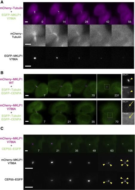
Figure 6.
Defects in the MKLP1–ARF6 interaction disintegrate the midbody. (A) Stills from the knockdown and rescue experiments with the GFP–MKLP1-V786A mutant performed in the HeLa cells expressing mCherry–tubulin. (B, C) Stills from the knockdown and rescue experiments with the mCherry–MKLP1-V786A mutant performed in the HeLa cells expressing GFP–tubulin and GFP–CENP-A (B) or GFP–CEP55 (C). Arrowheads in (B) indicate the intact midbody remnant with distinct signal for both MKLP1 and tubulin. Arrows indicate the residual MKLP1 signal in the regressing cell lacking a distinct tubulin signal. Arrowheads in (C) indicate the fragments from the disintegrating Flemming body. Bar, 5 μm.

