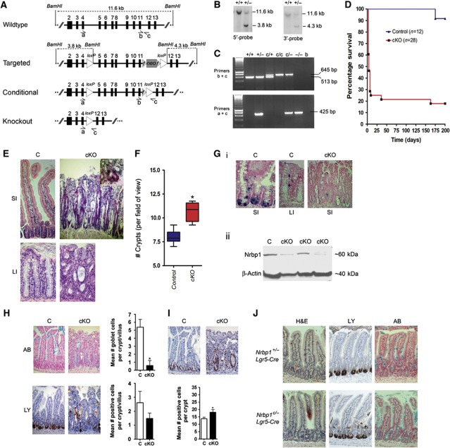
Figure 2.
Generation and characterisation of Nrbp1 knockout mice. (A) Schematic representation of the Nrbp1 locus, along with targeted, conditional and knockout Nrbp1 alleles. The PGK–neomycin selection cassette (neo) is flanked by Frt sites (striped triangles). Open triangles indicate LoxP sites for conditional deletion of exons 5–11. (B) Confirmation of correct targeting was performed by Southern blot analysis of BamHI-digested DNA; The wildtype band for both 5′- and 3′-probes was 11.6 kb and the targeted band was 3.8 and 4.3 kb, respectively. (C) Subsequent genotyping was performed by PCR, using either primers ‘b/c’ to detect the wild-type and conditional allele, or primers ‘a/c’ to detect the knock out allele. (D) Survival curve of Nrbp1flox, RosaCreERT2/+ (cKO) mice and controls after intraperitoneal dosing with 3 mg tamoxifen per day for 3 days. (E) H&E staining of the SI (small intestine) and LI (large intestine) from C (control; Nrbp1+/−RosaCreERT2/+ or Nrbp1flox/+RosaCreERT2/+) and cKO (Nrbp1flox/floxRosaCreERT2/+ or Nrbp1flox/−RosaCreERT2/+) mice at 5–7 days post tamoxifen treatment showing crypt elongation by primitive looking cells ( × 200 magnification). Inset: cKO intestines show evidence of crypt fission. (F) cKO mouse intestines had more crypts per field of view than C intestines at 4–5 days post dosing. (G) (i) ISH analysis using probe specific to Nrbp1 in the SI (small intestine) and LI (large intestine) of C mice (control; × 400 magnification) and the SI of cKO mice ( × 200 magnification) at 5 days post tamoxifen treatment, showing reduction of Nrbp1 expression levels in cKO tissue. (G) (ii) Western blot analysis of intestinal tissue harvested from C and cKO mice at 5 days post treatment with 1 mg tamoxifen for 4 days shows a reduction of Nrbp1 protein levels. β-Actin levels used as a control. (H) Histological and immunohistological analysis showing cKO mouse intestines had fewer goblet cells (as determined by Alcian blue staining) and an altered distribution of Paneth cells (as determined by lysozyme staining) compared to C mice at 4–5 days post dosing. *P<0.05. (I) cKO mice had more proliferating cells (as determined by Ki67 immunopositivity) than C intestines at 4–5 days post dosing. Data are represented as mean±s.d.; *P<0.05. (J) Histological and immunohistochemical analysis of the small intestines of 7–8-week-old Nrbp1−/cLgr5-EGFP-IRES-CreERT2+ and Nrbp1−/+Lgr5-EGFP-IRES-CreERT2+ control mice at 4–5 post dosing with 1 mg tamoxifen for 3 days. Data are represented as mean +/− s.d. (n=6 per genotype). Stem cell-specific deletion of Nrbp1 resulted in abnormal paneth cell localisation and granulisation, and abnormal goblet cell production ( × 200 magnification; haematoxylin and eosin, H&E; anti-lysozyme antibody, LY; Alcian blue, AB). **P<0.01, ***P<0.001.

