Figure 2.
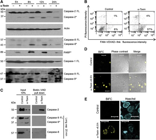
Caspase-2 is the initiator caspase during α-toxin-mediated apoptosis. (A) HeLa cells were treated with α-toxin (300 ng/ml) for various time points and the processing of caspases-2, -9, -8, -1 and PARP were monitored by immunoblots. Caspase-2 was detected by monoclonal antibody (clone 11b4; FL—full-length protein, *—processed form). (B) Flow cytometric analysis of caspase-2 activity. HeLa cells were incubated with or without α-toxin (300 ng/ml) for 24 h. Percentage of cells displaying caspase-2 activity is denoted. The cells were co-stained with PI to monitor membrane damage. (C) HeLa cells pretreated with biotin-VAD and treated with α-toxin as mentioned earlier. The activated caspases are precipitated as mentioned in the methods section. (D, E) BiFC of caspase-2 CARD. Hela cells transfected with venus–CARD constructs were treated with toxin, and BiFC (yellow) was measured under a confocal microscope. The nuclei are stained with Hoechst (blue) and the overlay is presented. Figure source data can be found with the Supplementary data.

