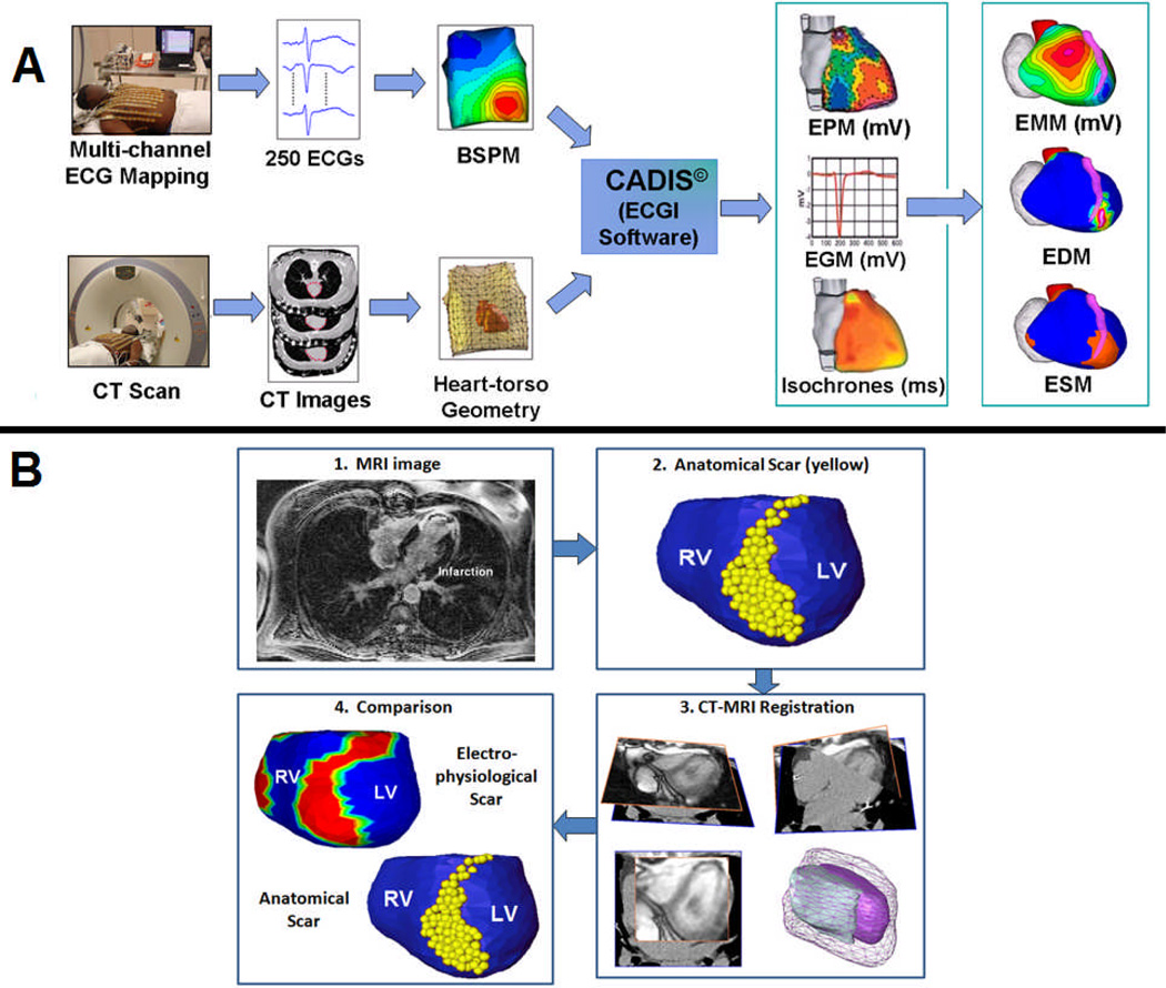
Figure 1.
A: The ECGI Procedure. Recorded body surface potentials and CT-imaged geometry are processed mathematically to obtain noninvasively potential maps (EPM), electrograms (EGM), and activation sequences (isochrones). From these data, electrogram magnitude maps (EMM), electrogram deflection maps (EDM) and electrical scar maps (ESM) are constructed (see text for details). ESM, defined by combining low magnitude potentials and EGMs with multiple deflections, is shown in red. B: Comparing electrical scar to anatomical scar. Anatomical scar is imaged with DE-MRI and, annotated (yellow dots) on the reconstructed cardiac geometry. The MRI image and CT from ECGI are co-registered to construct the anatomic scar map. A comparison of electrical (red) and anatomical (yellow) maps of scar on the anterior epicardial aspect of the interventricular septum is shown in B.4.

