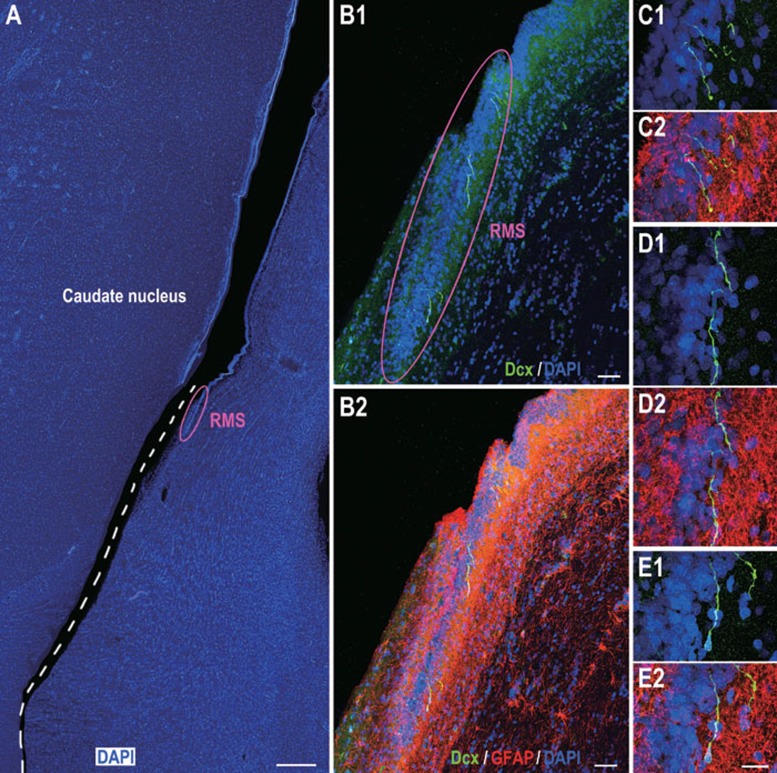
Figure 4.
Dcx/GFAP double immunostaining of adult human brain sections indicating Dcx+ cells in the RMS. (A) One representative section stained for DAPI showing the ventral SVZ and RMS. The section broke at the ventral SVZ (dashed line) during processing. (B1, B2) The photomicrographs showing GFAP+ astrocytic cells surrounding individual Dcx+ cells in the RMS. (C1-E2) Higher magnification of Dcx+ cells in B1 and B2. Scale bars represent 1 mm (A); 100 μm (B1, B2) and 20 μm (in E2 applies to C1-E2).

