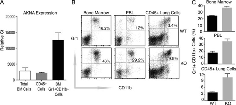
Figure 2.
Involvement of neutrophils in AKNA KO pathology. (A) The qRT-PCR results showing WT AKNA expression by murine bone marrow cells, CD45+ peripheral blood leukocytes (PBL) and pure bone marrow CD11b+Gr1+ neutrophils. Results are shown for three independent samples (n = 3) of each cell population ± SEM. P values < 0.05. (B) Comparative flow cytometry of CD11b+Gr1+ neutrophils levels between WT and AKNA KO mice in bone marrow, PBL and CD45+-gated lung cell suspensions. Values are presented (upper right quadrants) as the percentage of Gr1+ (y-axis) and CD11b+ (x-axis) cells. (C) Quantitative assessment of CD11b+Gr1+ neutrophils present in bone marrow, PBL and lung infiltrates of WT and KO mice. Bars indicate ± SEM (n = 6, P < 0.05).

