Abstract
In cysteine-rich peptides, diselenides can be used as a proxy for disulfide bridges, since the energetic preference for diselenide bonding over mixed selenium-sulfur bonds simplifies folding. Herein we report that an intramolecular diselenide bond efficiently catalyzes the oxidative folding of selenopeptide analogs of conotoxins, and serves as a reagentless method to substantially accelerate formation of various native disulfide bridging patterns.
Keywords: Diselenide, oxidative folding, kinetics, conotoxin, selenopeptide
Disulfide bonds are critical to stabilizing the native structure of many peptides and proteins. These cross-links are formed during oxidative folding, and machinery to assist formation of the correct cysteine pairings is used by all walks of life, from prokaryotes to humans.[1, 2] However, in the chemical synthesis of disulfide-containing biomolecules, alternative methods are employed to arrive at the native disulfide connectivity,[3] usually involving redox buffers and requiring peptide-specific optimization.[4]
Selenocysteine is often referred to as “nature’s 21st proteinogenic amino acid,” [5] and it can be readily incorporated into chemically synthesized peptides. Since diselenide bonds have greater thermodynamic stability than disulfide bonds, selenocysteine pairings can prevail over sequence-encoded folding information in short, cysteine-rich peptides.[6] By serving as a proxy for a disulfide bridge, selenocysteine incorporation into peptides may reduce the complexity of oxidative folding.[4,6–9] Consequently, diselenide bonds in peptides have been assumed to act as a fixed structural element during oxidative folding as a result of their high stability.
Diselenide bridges were incorporated into only a handful of cysteine-rich peptides including two-disulfide containing apamin[6,10] and α-conotoxins,[7,11] as well as three-disulfide bridged conotoxins.[4,8,12,13] Diselenides have also been useful to facilitate disulfide mapping.[12,14] While several groups have noted the enhanced kinetics of disulfide formation in selenopeptides,[8,11] to date, the rate enhancement has been assumed to be due to conformational effects.
Diselenide additives have been found to catalyze oxidative protein folding for a wide range of substrates.[15,16] With these additives, catalysis stems from rapid regeneration of the diselenide by reaction of the selenols formed in situ with atmospheric oxygen, even under (mildly) reducing conditions.[4,8] In contrast, thiols will not readily oxidize without a catalyst, such as a metal ion. Here we tested a hypothesis that if a diselenide can act as an intermolecular catalyst of cysteine oxidation, then disulfide-rich selenopeptides should be able to catalyze their own aerobic oxidative folding.
Conotoxins, which are derived from the venom of marine gastropods, represent an important target for improvements in oxidative folding efficiency. The venom of each species of Conus has a large and distinct array of disulfide-rich peptides, many of which are used as neuropharmacological tools.[17] While conotoxins have broad applicability, their oxidative folding is often quite challenging,[4,8,11,18,19] making conotoxins an attractive system for studying the mechanisms of oxidative folding.
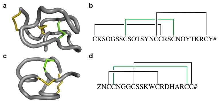
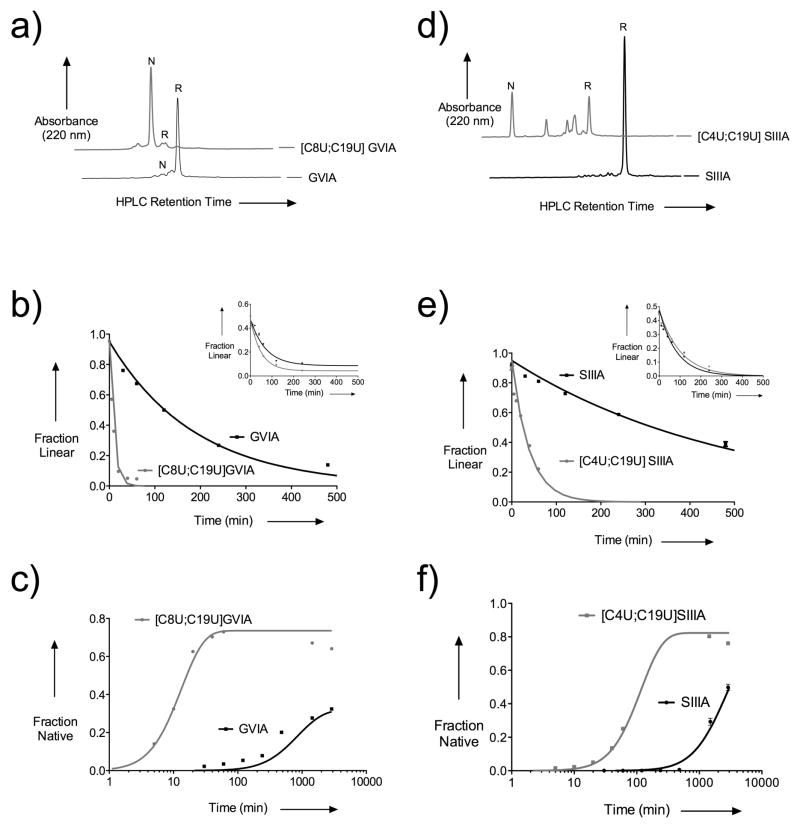
To examine whether diselenides enable the reagentless oxidative folding of cysteine-rich peptides, we selected previously characterized selenocysteine-containing analogs of ω-conotoxin GVIA and μ-conotoxin SIIIA (Figure 1).[4,8] Comparisons of the folding behavior for GVIA and [C8U;C19U] GVIA in Tris-buffered water are shown in Figure 2a–c. Both kox (the rate of formation of the first disulfide) and knative (the rate of appearance of the native connectivity) show significant enhancement in the presence of a diselenide, indicating its role in directing both oxidation and isomerization. To test the generality of this effect, similar experiments were also carried out using μ-conotoxin SIIIA and [C4U;C19U] SIIIA (Figure 2d–f). The significant increase in both kox (by approximately 12- to 20-fold) and knative (by approximately 34- to 175-fold) indicated that diselenides are efficient oxidative catalysts independent of the disulfide scaffold. Details of the kinetic analysis are presented in the Supporting Information.
Figure 1.
Folded products of peptides used in this study. a) shows the three-dimensional structure of GVIA (PDBID: 2CCO). b) shows the schematic representation of GVIA, where the bridge indicated in green was replaced with a diselenide in [C8U;C19U] GVIA. c) shows the three-dimensional structure of SIIIA (BMRB #20023). d) shows the schematic representation of SIIIA; the bridge indicated in green was replaced with selenocysteines in [C4U;C19U] SIIIA. Z indicates pyroglutamate, O indicates hydroxyproline, and # indicates C-terminal amidation.
Figure 2.
Folding kinetics of GVIA, SIIIA and their diselenide-containing analogs. a) and d) show HPLC chromatograms after 1 hour of folding, comparing GVIA and SIIIA to their diselenide-containing analogs, respectively. Full folding timecourses are available in the Supporting Information, Figure S4. b) and e) show kinetic traces of the disappearance of the starting material (bearing either six thiols or four thiols and a diselenide), with insets showing the same traces when the all-thiol and diselenide-containing peptides are folded in a 1:1 mixture. c) and f) show kinetic traces of the formation of natively-bridged peptide; equivalent traces using a linear timescale are shown in the Supporting Information, Figures S2 and S3.
The reagent-free formation of a disulfide bond by an intramolecular diselenide likely proceeds through three phases. First, the diselenide transfers an oxidizing equivalent to a pair of thiols to form a disulfide. Second, the selenol(ate) acts as a nucleophile to promote disulfide isomerization. Third, the selenol(ate)s are reoxidized by molecular oxygen in solution.
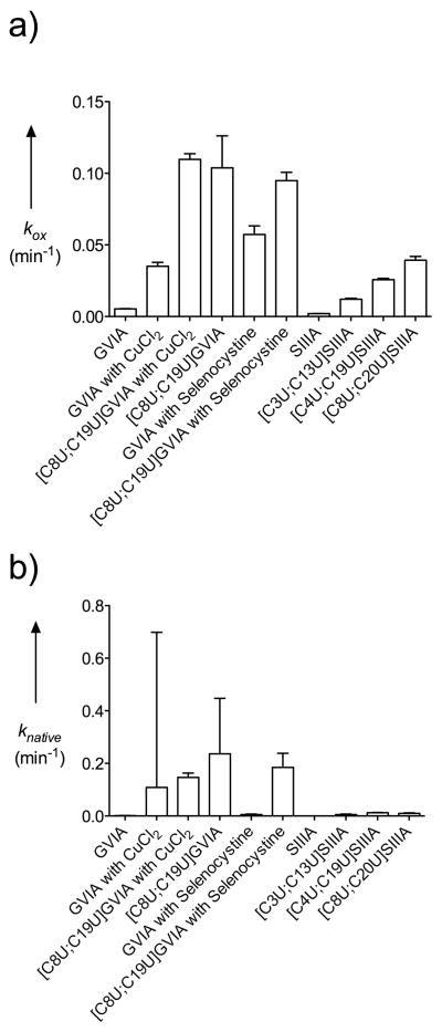
Both copper(II) and selenocystine are known to catalyze the oxidation of thiols to disulfides.[20,21] For both disulfide scaffolds considered, the addition of these oxidative catalysts afforded a substantial increase in the rate of oxidation (kox). Similarly, the rate of native formation (knative) was also enhanced, albeit to a much lower extent. However, for diselenide-containing peptides, neither oxidative catalyst provided a significant change in either the rate of oxidation or the rate of native formation (Figure 3). Interestingly, wild type GVIA folding in the presence of a selenocystine additive remains less efficient than that of reagentless folding with [C8U;C19U] GVIA.
Figure 3.
Rate constant comparison. a) shows the rate constants for initial oxidation, and b) shows the rate constants for the formation of the native connectivity. On each graph are compared GVIA, GVIA with 100 nM cupric chloride, [C8U;C19U] GVIA with 100 nM cupric chloride, [C8U;C19U] GVIA, GVIA with equimolar selenocystine, [C8U;C19U] GVIA with equimolar selenocystine, as well as variation due to the location of the diselenide within SIIIA.
The kinetic advantage of an intramolecular diselenide over added intermolecular diselenide in GVIA suggests a unimolecular mechanism. However, further analysis suggests that the reaction shows both inter- and intra-molecular character. On one hand, the initial oxidation of [C8U;C19U] GVIA appears concentration-independent across a 10-fold range in concentrations (Supporting Information, Figure S1); however, a 1:1 mixture of diselenide-containing peptide with its all-thiol counterpart suggests that intermolecular catalysis also contributes (Figure 2b and e, insets). This mixture of inter- and intra-molecular catalysis is particularly pronounced for SIIIA, where the initial oxidation step (kox) was slightly faster with added selenocystine (intermolecular catalysis), but isomerizations and subsequent oxidations leading ot the native disulfide connectivity (knative) were slightly faster with an intramolecular diselenide (Supporting Information, Table S1). Further discussion is provided in the Supporting Information.
To consider the role of diselenide placement within a peptide scaffold, we compared [C4U;C19U] SIIIA with [C3U;C13U] SIIIA and [C8U;C20U] SIIIA, which were chosen for this analysis because the reduced and folded forms are more readily separable by HPLC than the comparable analogs of GVIA. This data is shown in Figure 3 and in Supporting Information, Table S1, and demonstrates that the presence of a diselenide—regardless of its position in the peptide—greatly accelerates folding. While always beneficial, the position of the diselenide within the peptide does lead to some variation in the folding rate enhancement.
Thus, in addition to directing oxidative folding towards the native disulfide connectivity, intramolecular diselenide bridges also serve as catalysts for the formation of diuslfide bridges. A diselenide bridge in a peptide is therefore able to catalyze disulfide formation using dissolved oxygen as the stoichiometric oxidant, effectively enabling ‘reagentless’ oxidation. Using only molecular oxygen as the oxidant further simplifies the oxidative folding of cysteine-rich peptides. We envision the use of reagentless oxidation of selenopeptides, whether in solution or on-resin [4] will be effective, even for peptides that are difficult to synthesize.[19]
In the folding of selenopeptides, seleno-sulfide bonds are likely to be necessary intermediates for catalysis of intramolecular electron transfer reactions and isomerization steps. Examples of stable seleno-sulfide bonds are rare (Gowd, K.H., Blaise, K., Elmslie, K.S., Steiner, A.M., Olivera, B.M., and Bulaj, G., submitted). Non-native seleno-sulfide bonds have also been shown to exist in synthetic α-conotoxin and oxytocin analogs bearing a native pair of selenocysteines.[7,11,23] The high reactivity of selenocysteine is likely the reason why nature has selected disulfide bridges over diselenide bridges to form covalent crosslinks in polypeptides. To this end, our findings warrant a cautious approach towards exploring selenopeptides as drug leads. However, this does not preclude the added value of selenocysteine in the chemical synthesis of cysteine-rich peptides, as well as in manipulating oxidative folding pathways (Metanis, N. and Hilvert, D., submitted).
Our work shows that incorporation of diselenides into conotoxins may significantly improve their folding rates because the oxidative folding of the peptide is catalyzed by the peptide itself. In combination with previous reports, in which diselenides showed superior effects on simplifying and improving folding yields and disulfide mapping, this approach provides a significant advantage to chemical syntheses of cysteine-rich peptides. Since molecular biology techniques have already revealed thousands of bioactive peptides from plants, snails, spiders, scorpions and vertebrate animals, selenopeptide technologies will accelerate discoveries of novel research tools and biotherapeutics based on cysteine-rich peptides.
Experimental Section
Detailed methods are provided in the Supporting Information. Briefly, peptides were synthesized with standard Fmoc chemistry; all cysteine residues were trityl-protected, and all selenocysteine residues were 4-methoxybenzyl protected. Following cleavage from the resin, selenocysteine-containing peptides were treated with DTT. Peptides were then purified by HPLC prior to oxidative folding.
Unless otherwise indicated, all oxidations were air-mediated, using molecular oxygen as the only oxidant. Dried, reduced peptides were redissolved in 5% acetonitrile, 0.01% TFA. The peptide solution was then added to the folding mixture, which contained (final) 0.1 M Tris, pH 7.5, 0.1 mM EDTA, as well as any other folding additives for the specific condition. Folding reactions proceeded at 20 °C. Timepoints were analyzed by analytical HPLC.
HPLC peak integration was used to quantify the extent of peptide folding for each timepoint. This data was used to derive kinetic parameters using DynaFit. Details of the kinetic analysis are presented in the Supporting Information.
Supplementary Material
Footnotes
We thank Professors David Blair, David Goldenberg and Donald Hilvert for helpful discussions and comments on the manuscript. We also acknowledge the assistance of the Peptide Synthesis and Mass Spectrometry Core Facilities at the University of Utah. This work was supported by N.I.H. Program Project Grant GM 48677.
Supporting information for this article is available on the WWW under http://www.angewandte.org or from the author.
Contributor Information
Andrew M. Steiner, Department of Medicinal Chemistry, University of Utah, 421 Wakara Way, Suite 360, Salt Lake City, Utah 84108, (USA)
Kenneth J. Woycechowsky, Department of Chemistry, University of Utah, 315 S. 1400 E., Rm. 1320, Salt Lake City, Utah 84112
Prof. Dr. Baldomero M. Olivera, Department of Biology, University of Utah, 257 S. 1400 E., Rm. 115, Salt Lake City, Utah 84112
Prof. Dr. Grzegorz Bulaj, Email: bulaj@pharm.utah.edu, Department of Medicinal Chemistry, University of Utah, 421 Wakara Way, Suite 360, Salt Lake City, Utah 84108, (USA); Fax: (+1) 801-581-7087
References
- 1.Nakamoto H, Bardwell JC. Biochem Biophys Acta. 2004;1694:111. doi: 10.1016/j.bbamcr.2004.02.012. [DOI] [PubMed] [Google Scholar]
- 2.Ellgaard L, Ruddock LW. EMBO Rep. 2005;6:28. doi: 10.1038/sj.embor.7400311. [DOI] [PMC free article] [PubMed] [Google Scholar]
- 3.Annis I, Hargittai B, Barany G. Methods Enzymol. 1997;289:198. doi: 10.1016/s0076-6879(97)89049-0. [DOI] [PubMed] [Google Scholar]
- 4.Steiner AM, Bulaj G. J Pep Sci. 2011;17:1. doi: 10.1002/psc.1283. [DOI] [PubMed] [Google Scholar]
- 5.Allmang C, Wurth L, Krol A. Biochim Biophys Acta. 2009;1790:1415. doi: 10.1016/j.bbagen.2009.03.003. [DOI] [PubMed] [Google Scholar]
- 6.Fiori S, Pegoraro S, Rudolph-Böhner S, Cramer J, Moroder L. Biopolymers. 2000;53:550. doi: 10.1002/(SICI)1097-0282(200006)53:7<550::AID-BIP3>3.0.CO;2-O. [DOI] [PubMed] [Google Scholar]
- 7.Armishaw CJ, Daly NL, Nevin ST, Adams DJ, Craik DJ, Alewood PF. J Biol Chem. 2006;281:14136. doi: 10.1074/jbc.M512419200. [DOI] [PubMed] [Google Scholar]
- 8.Gowd KH, Yarotskyy V, Elmslie KS, Skalicky JJ, Olivera BM, Bulaj G. Biochemistry. 2010;49:2741. doi: 10.1021/bi902137c. [DOI] [PMC free article] [PubMed] [Google Scholar]
- 9.Walewska A, Jaśkiewicz A, Bulaj G, Rolka K. Chem Biol Drug Des. 2011;77:93. doi: 10.1111/j.1747-0285.2010.01046.x. [DOI] [PubMed] [Google Scholar]
- 10.Pegoraro S, Fiori S, Cramer J, Rudolph-Böhner S, Moroder L. Protein Sci. 1999;8:1605. doi: 10.1110/ps.8.8.1605. [DOI] [PMC free article] [PubMed] [Google Scholar]
- 11.Muttenthaler M, Nevin ST, Grishin AA, Ngo ST, Choy PT, Daly NL, Hu SH, Armishaw CJ, Wang CI, Lewis RJ, Martin JL, Noakes PG, Craik DJ, Adams DJ, Alewood PF. J Am Chem Soc. 2010;132:3514. doi: 10.1021/ja910602h. [DOI] [PubMed] [Google Scholar]
- 12.a) Walewska A, Zhang MM, Skalicky JJ, Yoshikami D, Olivera BM, Bulaj G. Angew Chem Int Ed Engl. 2009;48:2221. doi: 10.1002/anie.200806085. [DOI] [PMC free article] [PubMed] [Google Scholar]; b) Walewska A, Zhang MM, Skalicky JJ, Yoshikami D, Olivera BM, Bulaj G. Angew Chem. 2009;121:2255. doi: 10.1002/anie.200806085. [DOI] [PMC free article] [PubMed] [Google Scholar]
- 13.Han TS, Zhang MM, Gowd KH, Walewska A, Yoshikami D, Olivera BM, Bulaj G. ACS Med Chem Lett. 2010;1:140. doi: 10.1021/ml900017q. [DOI] [PMC free article] [PubMed] [Google Scholar]
- 14.Mobli M, de Araújo AD, Lambert LK, Pierens GK, Windley MJ, Nicholson GM, Alewood PF, King GF. Angew Chem Int Ed Engl. 2009;48:9312. doi: 10.1002/anie.200905206. [DOI] [PubMed] [Google Scholar]; b) Mobli M, de Araújo AD, Lambert LK, Pierens GK, Windley MJ, Nicholson GM, Alewood PF, King GF. Angew Chem. 2009;121:9476. doi: 10.1002/anie.200905206. [DOI] [PubMed] [Google Scholar]
- 15.Beld J, Woycechowsky KJ, Hilvert D. Biochemistry. 2007;46:5382. doi: 10.1021/bi700124p. [DOI] [PubMed] [Google Scholar]
- 16.Beld J, Woycechowsky KJ, Hilvert D. J Biotechnol. 2010;150:481. doi: 10.1016/j.jbiotec.2010.09.956. [DOI] [PubMed] [Google Scholar]
- 17.Norton RS, Olivera BM. Toxicon. 2006;48:780. doi: 10.1016/j.toxicon.2006.07.022. [DOI] [PubMed] [Google Scholar]
- 18.Han TS, Zhang MM, Walewska A, Gruszczynski P, Robertson CR, Cheatham TE, Yoshikami D, Olivera BM, Bulaj G. ChemMedChem. 2009;4:406. doi: 10.1002/cmdc.200800292. [DOI] [PMC free article] [PubMed] [Google Scholar]
- 19.a) de Araujo AD, Callaghan B, Nevin ST, Daly NL, Craik DJ, Moretta M, Hopping G, Christie MJ, Adams DJ, Alewood PF. Angew Chem Int Ed Engl. 2011;50:6527. doi: 10.1002/anie.201101642. [DOI] [PubMed] [Google Scholar]; b) de Araujo AD, Callaghan B, Nevin ST, Daly NL, Craik DJ, Moretta M, Hopping G, Christie MJ, Adams DJ, Alewood PF. Angew Chem. 2011;123:6657. doi: 10.1002/anie.201101642. [DOI] [PubMed] [Google Scholar]
- 20.Kachur AV, Koch CJ, Biaglow JE. Free Rad Res. 1999;31:23. doi: 10.1080/10715769900300571. [DOI] [PubMed] [Google Scholar]
- 21.Beld J, Woycechowsky KJ, Hilvert D. Biochemistry. 2008;47:6985. doi: 10.1021/bi8008906. [DOI] [PubMed] [Google Scholar]
- 22.Mullenbach GT, Tabrizi A, Irvine BD, Bell GI, Hallewell RA. Nucleic Acids Res. 1987;15:5484. doi: 10.1093/nar/15.13.5484. [DOI] [PMC free article] [PubMed] [Google Scholar]
- 23.Muttenthaler M, Andersson A, Araujo AD, Dekan Z, Lewis RJ, Alewood PF. J Med Chem. 2010;53:8585. doi: 10.1021/jm100989w. [DOI] [PubMed] [Google Scholar]
- 24.Swain CG. J Am Chem Soc. 1944;66:1696. [Google Scholar]
Associated Data
This section collects any data citations, data availability statements, or supplementary materials included in this article.


