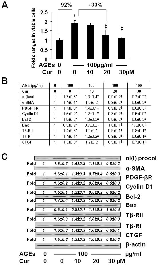
Figure 2. Curcumin eliminated the effects of AGEs on the induction of HSC activation.

Serum-starved HSCs were treated with or without AGEs-BSA at 100 μg/ml plus or minus curcumin at indicated concentrations in serum-depleted media for 24 hr. (A) Cell proliferation was determined by MTS assays. Results were expressed as fold changes in the number of viable cells, compared with the untreated control (mean± s. d., n=3). *p<0.05 vs. the untreated control (the 1st column); ‡p<0.05 vs. cells treated with AGEs alone (the 2nd column). The percentage in difference was calculated in the formula: [(# in target HSCs − # in compared HSCs)/# in compared HSCs] x 100%. (B) real-time PCR analyses. Values were presented as mRNA fold changes (mean ± s. d., n=3). *p<0.05 vs. the untreated control; ‡p<0.05 vs. cells treated with AGEs alone. (C) Western blotting analyses. Representatives were from three independent experiments. β-actin was used as an internal control for equal loading. Italic numbers beneath blots were fold changes (means ± s. d., n=3) in the densities of the bands compared with the control without treatment in the blot, after normalization with the internal invariable control.
