
Figure 7. The activation of PPARγ eliminated the inhibitory effect of AGEs and stimulated gene expression of AGE-R1 in HSCs in vitro.
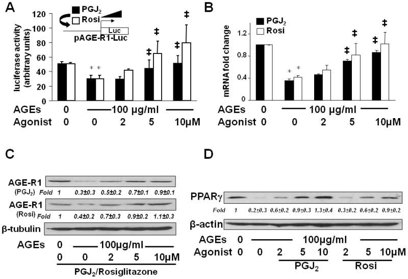
(A) HSCs were transfected with the AGE-R1 promoter luciferase reporter plasmid pAGE-R1-Luc. After recovery, cells were serum-starved for 4 hr prior to the treatment with or without AGEs (100 μg/ml) in the presence of PGJ2, or rosiglitazone (Rosi), at 0–10 μM in serum-depleted media for 24 hr. Luciferase activity assays were conducted (n=6). *p<0.05 vs. cells with no treatment (the corresponding 1st column). ‡p<0.05 vs. cells treated with AGEs alone (the corresponding 2nd column). The floating schema denoted pAGE-R1-Luc in use for transfection and the application of PGJ2, or Rosi, to the system.
(B, C & D) Serum-starved HSCs were stimulated with or without AGEs (100 μg/ml) in the presence of PGJ2, or Rosi, at 0–10 μM in serum-depleted media for 24 hr. Total RNA and whole cell extracts were prepared. (B). real-time PCR assays. Values were presented as mRNA fold changes (mean ± s. d., n=3). *p<0.05 vs. cells with no treatment (the corresponding 1st column). ‡p<0.05 vs. cells treated with AGEs alone (the corresponding 2nd column). (C & D) Western blotting analyses. β-tubulin, or β-actin, was used as an internal control for equal loading. Representatives were from three independent experiments. Italic numbers beneath blots were fold changes (mean ± s. d., n=3) in the densities of the bands compared with the control without treatment in the blot, after normalization with the internal invariable control.