Figure 4.
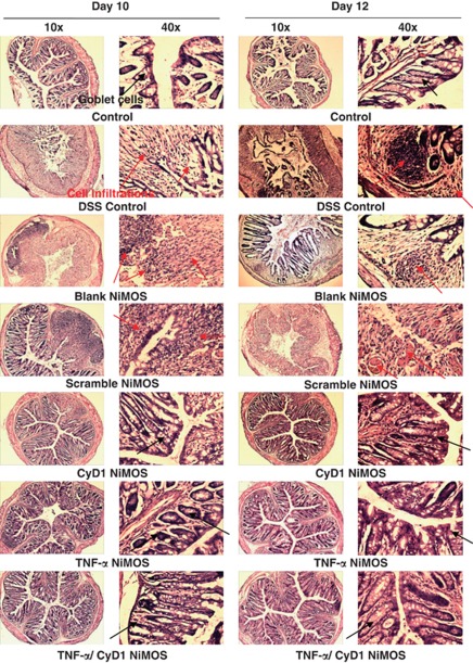
Microscopic evaluation of colonic tissue histopathology. Bright-field images of haematoxylin and eosin stained sections of the colon harvested from each control and test group. Images are shown at magnifications of × 10 and × 40 from tissue cryosections obtained on day 10 and day 12 of the study. Sections from the first control group show normal and healthy colon tissue. Intestinal tissues from the dextran sulfate sodium (DSS) control group, the group treated with blank and scrambled short interfering RNA (siRNA) nanoparticles-in-microsphere oral system (NiMOS) showed a severe infiltration of white blood cells, abnormal mucosal structure, and a certain degree of goblet cell depletion. Tissue from the group receiving tumor necrosis factor-α (TNF-α), cyclin D1 (CyD1), or combined TNF/CyD1 silencing NiMOS showed signs of regeneration and exhibited a tissue architecture more closely resembling that of healthy tissue in the normal control group. Occurrence of goblet cells is indicated by red arrows; cell infiltration and abnormal tissue histology is indicated by black arrows.

