FIGURE 4.
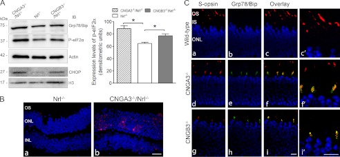
Enhanced expression of the ER stress marker proteins in CNGA3−/−/Nrl−/− and CNGB3−/−/Nrl−/− retinas. The expression levels of Grp78/Bip, phospho-eIF2α, and CHOP were examined in CNGA3−/−/Nrl−/−, CNGB3−/−/Nrl−/−, and Nrl−/− mice at P30. A, left panel: shown are representative images of the Western blot detections. Total retinal protein lysate was used for detection of Grp78/Bip and phospho-eIF2α (actin was used as a loading control, upper three panels), and retinal nuclear preparation was used for detection of CHOP. (H3 was used as a loading control, lower two panels.) Right panel: densitometric analysis of the relative expression levels of phospho-eIF2α in CNGA3−/−/Nrl−/−, CNGB3−/−/Nrl−/−, and Nrl−/− retinas. Data are represented as means ± S.E. of measurements from three to four independent experiments using retinas from four to five mice. Unpaired Student's t test was used for determination of the significance (*, p < 0.05). B, CHOP activation in CNG channel-deficient retina. Shown are images of CHOP staining on retinal sections of Nrl−/− (a) and CNGA3−/−/Nrl−/− (b) mice. C, co-localization of Grp78/Bip with S-opsin in CNG channel-deficient retina. Co-labeling was performed using rabbit anti-Grp78/Bip and goat anti-S-opsin antibodies. Shown are images of the co-labeling on retinal sections of WT (a–c), CNGA3−/− (d–f), and CNGB3−/− (g–i) mice with higher magnification images alongside (c′, f′, i′). IS, inner segment; ONL, outer nuclear layer; IB, immunoblot. Scale bar, 20 μm.

