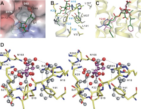
FIGURE 4.
Overall structure of the Y248A CrtM–ZA-A complex. A, electrostatic surface representation of Y248A CrtM binding site with ZA-A (green). Here, the S2 site is empty. B, superimposition of apo-CrtM (gray) and Y248A CrtM–ZA-A (yellow) showing the local conformational changes of the S1 site upon binding to ZA-A. C, ZA-A binds to the Y248A CrtM active site. If present, the C-6 acyl group would crash into Tyr248 (in slate). D, close-up view of the detailed interactions of ZA-A central core (magenta carbons) with Y248A CrtM shown in stereo. The ZA-A molecule is shown as a ball-and-stick representation. The important residues are labeled. Potential hydrogen bonds between the side chains of Y248A CrtM and ZA-A are shown as black dashes.

