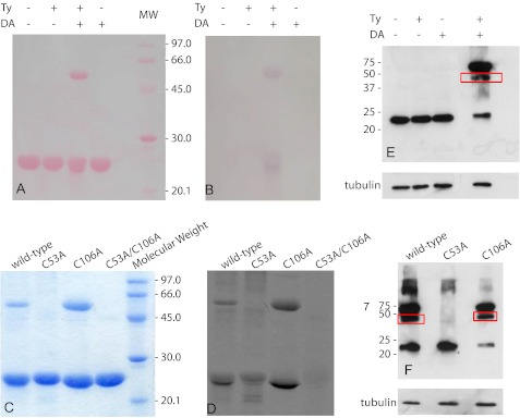
FIGURE 1.
Identification of DAQ adducts formed on DJ-1. WT DJ-1 samples reacted with DA and/or Ty were run on an SDS-polyacrylamide gel. A, Ponceau S staining of the nitrocellulose membrane on which the SDS-polyacrylamide gel was transferred. B, redox-cycling staining assay run on the nitrocellulose membrane reported in A. C, SDS-polyacrylamide gel of WT DJ-1 and mutant protein samples previously exposed to 14C-DA, in the presence of Ty, in a 3:1 cysteine/DA ratio. D, autoradiography obtained from the SDS-polyacrylamide gel reported in C. E, Western blot analysis performed using a monoclonal DJ-1 antibody on endogenous DJ-1 from cell lysates after treatment with DA and/or Ty. F, Western blot analysis performed using a V5 antibody on transfected cells containing WT DJ-1, C53A, or C106A mutants. Cells were lysed and treated with DA and/or Ty before Western blot analysis. Mouse anti-β-tubulin was used as an internal control.

