FIGURE 5.
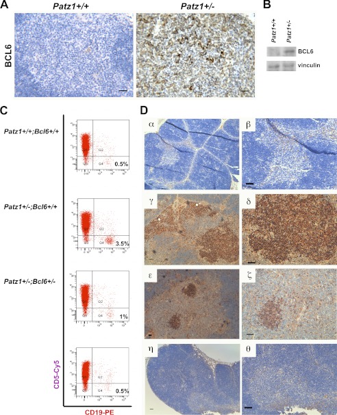
Key role of BCL6 in thymus B cell expansion of Patz1 knock-out mice. A, immunohistochemical staining of BCL6 in representative thymus samples from Patz1+/+ (left panel) and Patz1+/− (right panel) mice. Scale bar, 100 μm. B, Western blot analysis for BCL6 expression in a pool of three Patz1+/+ and three Patz1+/− thymi. C, flow cytometry of representative thymi from Patz1+/+;Bcl6+/+, Patz1+/−;Bcl6+/+, and Patz1+/−;Bcl6+/− mice. The thymocytes were double-stained for specific B (CD19) and T cell (CD5) surface antigens. For each analysis, 10,000 events were counted. The relative percentage of CD19+ cells (B lymphocytes) was indicated in the right-bottom corner of each dot plot. CD19-PE, CD19-Phycoerythrin staining. D, immunohistochemical analysis of the thymi shown in C to detect B cells (stained with anti-B220). Only scattered positive cells were detected in Patz1+/+;Bcl6+/+ (α,β) and most Patz1+/−;Bcl6+/− thymi (η,θ). Large focal hyperplasias of B cells were detected in Patz1+/−;Bcl6+/+ (γ, δ) thymi, and small focal positivity was detected in some Patz1+/−;Bcl6+/− thymi (ϵ, ζ). Scale bars, 100 μm.

