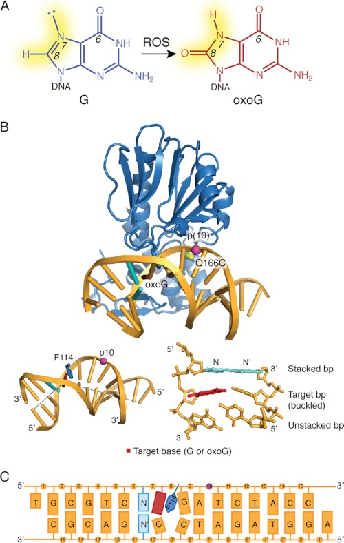
FIGURE 1.
A, formation of oxoG from guanine (G) by reactive oxygen species (ROS) is shown. The changes in atom substituents at N7 and C8 are highlighted in yellow. B, the structure of MutM cross-linked to an intrahelical oxoG (PDB code 3GP1) is shown. The cross-linking sites on the protein and DNA are as indicated. A view of the base-stacking to the target base is shown in the bottom right (with the stacked base pair shown in cyan); insertion of Phe114 into the duplex and the resulting buckle in the DNA (with gray lines marking the helical axis flanking the target base) is shown at the bottom left. C, shown is a schematic diagram of the DNA used in this study. The target base, shown in red, was either G or oxoG; base N is the 5′-stacking neighbor fg and was varied between A, T, and G. The modified phosphate group is shown in purple.

