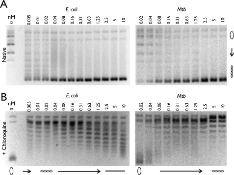
FIGURE 5.
E. coli and M. tuberculosis negative supercoiling holoenzyme assays. A and B, a portion of each sample was run on a 1% agarose gel in the absence (A) and presence (B) of 3 μg/ml chloroquine. Protein concentrations are listed in nm holoenzyme, and DNA topoisomers are labeled with graphic representations across the bottom of the chloroquine gel. Relaxed and supercoiled DNA species are labeled with graphic representations on the right of the native gel. Mtb, M. tuberculosis.

