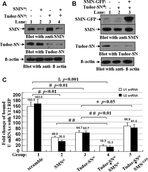
FIGURE 8.
Ectopically expressed SMN restored the reduced U snRNP assembly caused by depletion of endogenous Tudor-SN. A and B, HeLa cells were transfected with the Tudor-SN siRNA and SMN siRNA (A) or mammalian expression plasmids containing full-length SMN tagged with GFP epitope (SMN-GFP) as indicated (B). The total cell lysates of different samples were blotted with anti-SMN (upper panel), anti-Tudor-SN (middle panel), or anti-β-actin antibody (lower panel) to detect the protein level of corresponding proteins. C, total lysates of different samples were immunoprecipitated with Y12 Dynabeads or anti-IgG Dynabeads as control. The bound RNAs were isolated and reverse-transcribed to cDNA with random hexamer primers, and we then performed the quantitative real time PCR assay to detect the relative fold changes of precipitated U1 and U2 snRNA. The RIP fold changes were analyzed with analysis of variance. Significant difference was indicated as follows: &, <0.001; #, p < 0.01 versus nontreatment group (n = 3), *, p < 0.05; # #, p < 0.01 versus Tudor-SNsi or SMNsi group (n = 3).

