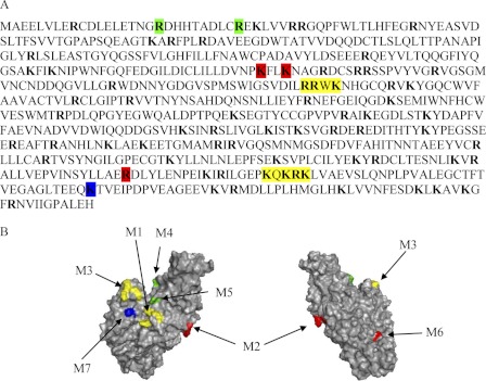
FIGURE 1.
Identification of potential heparin binding sites on the TG2 sequence. A, shown is the amino acid sequence of TG2. The basic residues Arg and Lys are in bold, and the colored boxes indicate residues investigated by mutagenesis. B, the structure of TG2 (PDB code 3LY6) shows the surface localization of residues corresponding to TG2 mutants M1 and M3 (yellow), M2 and M6 (red), M4 and M5 (green), and M7 (blue). The drawings were prepared with PyMOL.

