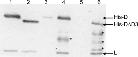
FIGURE 9.
Co-purification of endogenous subunit L with His-tagged subunit D or subunit DΔD3 expressed within M. acetivorans. Shown is a Western blot of a 15% SDS-polyacrylamide gel with anti-D-L antibodies. Lane 1, recombinant D-L(His) heterodimer (50 ng); lane 2, recombinant DΔD3-L(His) heterodimer (150 ng); lane 3, imidazole eluate from a Ni2+-agarose column loaded with cell lysate of DJL30 grown in the absence of tetracycline; lane 4, imidazole eluate from a Ni2+-agarose column loaded with cell lysate of DJL30 grown in the presence of tetracycline; lane 5, imidazole eluate from a Ni2+-agarose column loaded with cell lysate of DJL31 grown in the absence of tetracycline; lane 6, imidazole eluate from a Ni2+-agarose column loaded with cell lysate of DJL31 grown in the presence of tetracycline. The asterisks indicate subunit His-D degradation products.

