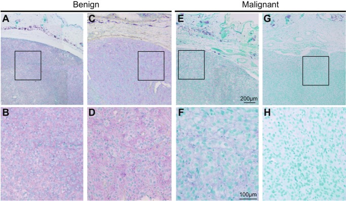
Figure 3.
Immunohistochemical analysis of Gng2 protein expression in murine tumors. Results of immunohistochemistry for Gng2 protein in benign tumors (A-D) and malignant melanomas (E-H) from RET-mice of line 304/B6 are presented. Bottom panels are shown as higher magnification of top panels. Staining of Gng2 protein and the nucleus are presented as purple and green, respectively.

