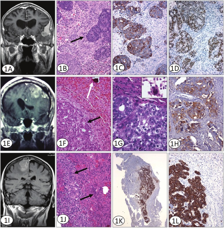
Figure 1.
A. Coronal T1 weighted MRI with contrast shows enhancement of the pterional meningioma, with dural tail sign superiorly and metastatic lesion involving the anteromedial part of the tumor. Although radiographically a collision tumor could be considered, histologically islands of metastatic adenocarcinoma surrounded entirely by meningioma are seen. B. H&E stained sections illustrate metastatic colorectal adenocarcinoma (solid arrow) within otherwise typical meningioma. Original magnification 200x. C. Immunohistochemistry for Cytokeratin 20 supports a colorectal origin for the metastatic component defining solid areas and islands of immunoreactive tumor. Cytokeratin 20 immunoreacted sections. Original magnification 200x. D. Immunohistochemistry for Cdx-2 confirms the origin of the tumor and defines solid areas and islands of immunoreactive tumor. Cdx-2 immunoreacted sections. Original magnification 200x. E. MRI revealed a large, 6cm left frontal mass containing blood adjacent to a prominent area of calvarial hyperostosis. Both intra and extra-axial components were identified. The tumor was creating a mass effect with surrounding vasogenic edema. F and G. H&E stained sections illustrate two morphologically distinct areas; an epithelial, glandular component (black arrow) and a second area with a syncytial pattern and a well defined psammoma body (white arrow) is identified. At higher magnification the larger nuclei and prominent nucleoli of the prostatic adenocarcinoma are evident. The inset shows a well-formed meningothelial whorl seen on smear preparation. (Original magnifications 100x and 400x). H. The metastatic adenocarcinoma component of the tumor is prostate specific antibody immunoreactive. Immunohistochemistry for PSAP Original magnification 200x. I. MRI revealed a dural-based, 2.4 x2.6cm lesion located in the area of the right frontal cortex with some vasogenic edema. The lesion displayed some heterogeneity with apparent discrete areas within the tumor which differed from the larger surrounding tumor. J. H&E stained sections illustrate metastatic prostate adenocarcinoma (solid arrow) within meningioma. Original magnification 200x. K and L. The prostate adenocarcinoma component of the tumor demonstrates a discretely positive cytokeratin Cam 5.2 reaction. Immunohistochemistry for Cytokeratin Cam 5.2. Original magnification 100x and 200x.

