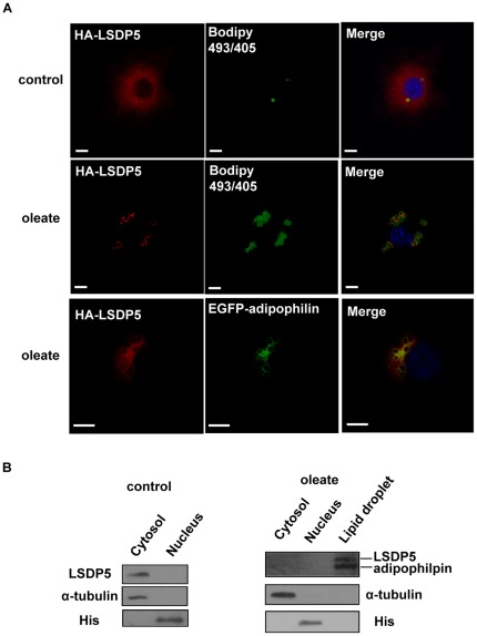
Figure 1. LSDP5 was recruited to lipid droplets.
(A) AML12 cells were transiently transfected with HA-tagged LSDP5 and incubated with BSA (control) or oleate (100 µM) overnight. The cells were stained with an anti-HA antibody with BODIPY 493/503 for visualizing lipids (green) and Hoechst 33258 for visualizing nuclei (blue). Left panels show the immunofluorescent signal (red), middle panels showed BODIPY staining (green), and right panels show the merged images. HA-LSDP5 exhibited a steady-state faint cytoplasmic staining and decorated lipid droplets after incubating the cells in oleate-rich medium. AML12 cells were co-transfected with HA-LSDP5 and EGFP-adipophilin. The cells were incubated with a mouse anti-HA antibody (primary antibody) and a Cy3-conjugated anti-mouse antibody (secondary antibody). The samples were detected by fluorescence microscopy (Olympus, Temecula, CA). The results show the co-localization of LSDP5 with adipophilin, a lipid droplet-targeted protein (last row). Scale bar = 5 µm. (B) LSDP5 was enriched in lipid droplet fractions. α-tubulin, a cytosol marker; His, a nucleus marker; and adipophilin, a lipid droplet marker. 5 mg of each fraction was loaded for immunoblot analysis.

