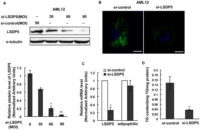
Figure 4. LSDP5 deficiency inhibited TG accumulation in AML12 cells.
AML12 cells were infected with an adenovirus carrying LSDP5 siRNA for 24 h and then incubated with 200 µM oleate for another 24 h. (A) Western blotting revealed that the adenovirus (si-LSDP5) at a multiplicity of infection (MOI) of 90 successfully silenced LSDP5 in AML12 cells (>95% knock-down). Expression levels of LSDP5 are expressed as a ratio to α-tubulin (representative of three experiments). Data are presented as the mean±SEM. * P<0.05, ** P<0.01 (Dunnett’s post hoc test following a one-way ANOVA). (B) BODIPY staining of AML12 cells expressing control siRNA (left) or LSDP5 siRNA (right). Scale bar = 15 µm. (C) The mRNA levels of LSDP5 and adipophilin were assessed with real-time PCR. The relative mRNA level in AML12 cells infected with an adenovirus containing control siRNA was designated as 1.0. Data are presented as the mean±SEM (n = 4), * P<0.05. (D) A lower concentration of TGs was detected in si-LSDP5 cells, compared with control cells. Data are presented as the mean±SEM (n = 5), * P<0.05 (paired Student’s t test).

