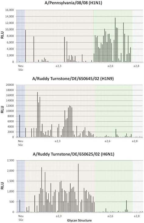
Figure 3. Glycan binding analysis of wild bird avian or human influenza viruses.
Influenza viruses were propagated in Madin-Darby kidney cells, purified on a 25% sucrose cushion by ultracentrifugation, and labeled with Alexa488 before being applied to the microarray. The data was organized based on Neu5GC, α2,3 SA, α2,6 SA and α2,8 SA glycan structures and represented by different color schemes. Glycan microarray binding analysis was performed by Core H of the Consortium for Functional Glycomics. A) A/Ruddy Turnstone/DE/1171/02 (H1N9), B) A/Ruddy Turnstone/DE/892/02 (H6N1), C) A/Pennsylvania/08/2008 (H1N1).

