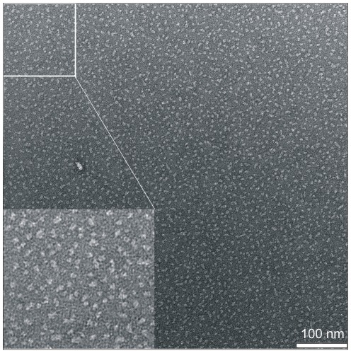
Figure 8. Negative-stain TEM of single purified synaptogyrin 1 protein in the presence of the detergent DDM.
The homogeneity of the purified protein is reflected in the electron micrograph. Synaptogyrin particles have a diameter of around 5–6 nm. The scale bar represents 100 nm. Magnifications are shown.

