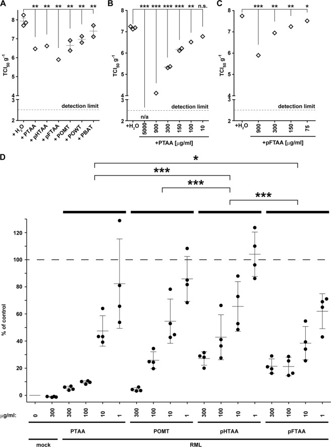
FIGURE 2.
LCPs reduce prion infectivity and the amount of prion aggregates in RML6 brain homogenates. A, SCEPA of RML6 homogenates mixed with the different LCPs at a concentration of 300 μg/ml. B and C, SCEPA of RML6 homogenates mixed with increasing concentrations of PTAA (B) and pFTAA (C). Noninfected (mock) and RML6-infected brain homogenates were incubated with either PTAA or pFTAA, and infectivity was determined on 17,000 cells in the SCEPA. Each data point represents the infectivity of one sample determined by serial 10-fold dilution steps from 10−4 to 10−8 on a 96-well plate. Data are shown as means ± S.E. p values represent statistical difference between polythiophene-treated and nontreated RML homogenates and were calculated with a Mantel-Haenszel χ2 test (supplemental Table S1). Detection limit, theoretical infectivity based on the observation of false positives at concentrations between 10−2 and 10−3 of noninfectious inoculum (mock). ***, p < 0.001; **, p < 0.01; *, p < 0.05. n/a, no positive wells were observed with this sample between concentrations ranging from 10−3 to 10−8. ns, nonsignificant, p > 0.05. D, MPA assay was used to quantify prion aggregates in RML6 brain homogenates treated with increasing concentrations of PTAA, POMT, pHTAA, or pFTAA. Each sample was analyzed in technical quadruplicates (circles) and is represented as the mean ± S.D. All data were corrected for the negative control (mock, CD1 brain homogenate) and represented as relative light units normalized for RML6 prions. Statistical differences were computed to compare LCP-treated RML6 to the control (nontreated RML6) (supplemental Table S1) or groups of four concentrations of each polythiophene (supplemental Table S6).

