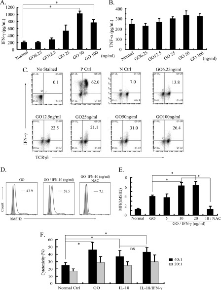
FIGURE 5.
IFN-γ produced in oxidative stress promotes ectopic expression of hMSH2 and lysis of RCC cells by γδ T cells. A and B, the secretion of IFN-γ or TNF-α by GO-stimulated γδ T cells was measured by ELISA (mean ± S.D.; n = 5 independent experiments). *, p < 0.05. C, intracellular staining detection of IFN-γ produced by GO-stimulated γδ T cells. γδ T cells incubated with 20 ng/ml phorbol 12-myristate-13-acetate (PMA), 0.5 μg/ml ionomycin, and 3 μg/ml brefeldin A for 4 h acted as the positive control. Normally cultured γδ T cells incubated with 3 μg/ml brefeldin A for 4 h are shown as the negative control. D, surface expression of hMSH2 on GO-stimulated G401 cells in the presence or absence of exogenous IFN-γ (10 ng/ml) followed by treatment with or without NAC (5 mm). Surface expression of hMSH2 on G401 cells is depicted with black lines, and the isotype controls are displayed in solid gray. E, mean fluorescence intensity (MFI) of ectopic expression of hMSH2 treated by GO with or without exogenous IFN-γ is shown. Error bar, S.D. n = 3 independent experiments. *, p < 0.05. ns, not significant. F, γδ T cell-mediated lysis of G401 cells and G401 cells treated with GO (100 ng/ml), IL-18 (100 ng/ml), or IL-18 (100 ng/ml) plus IFN-γ (10 ng/ml) at E:T ratio 20:1 or 40:1. Error bars, S.D. n = 3 independent experiments. *, p < 0.05.

