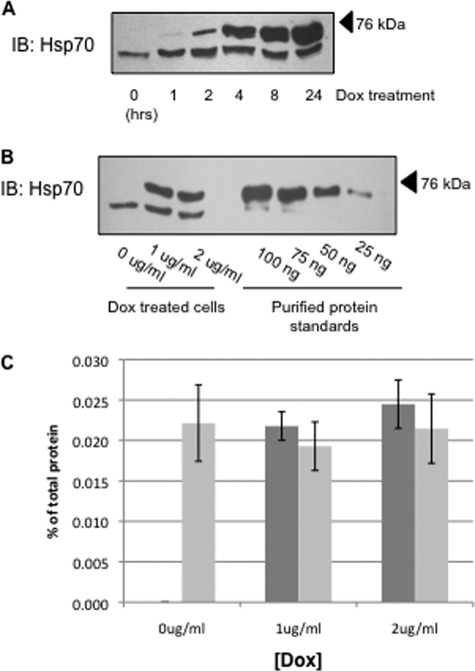
FIGURE 1.
Overexpression of Hsp70 in MDCK cell lysates. A, MDCK cells were treated with 1 μg/ml Dox for indicated time periods, and Hsp70 was detected in cell lysates by immunoblot (IB). The lower bands and upper bands represent the endogenous Hsp70 protein and the Myc/His-tagged construct of Hsp70 protein, respectively. B, Hsp70 expression was determined by immunoblot after a 24-h incubation with the indicated amount of Dox. This immunoreactivity was compared with that of purified Hsp70 protein to quantify Hsp70 expression. C, densitometric analysis of bands from five independent Hsp70 immunoblots quantifying endogenous (light bars) and overexpressed (dark bars) chaperone. The error bars indicate S.E.

