FIGURE 3.
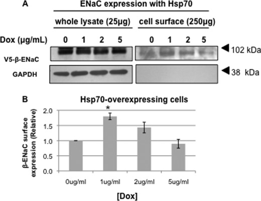
Hsp70 overexpression increases the surface expression of ENaC in MDCK cells. MDCK cells were treated with the indicated amount of Dox for 24 h. β-ENaC at the apical surface was detected by surface biotinylation as described under “Experimental Procedures.” A, representative immunoblots for β-ENaC (using an antibody to the V5 epitope tag) of whole cell lysates and NeutrAvidin-precipitated proteins are shown. Immunoblots for GAPDH were used as a control for protein loading (whole cell lysates) and to ensure that the membrane-impermeant biotin did not label intracellular proteins (NeutrAvidin precipitates). B, densitometric quantification of immunoblots from three independent biotinylation experiments. The error bars indicate S.E. *, p = 0.004 versus 0 μg/ml Dox treatment (ANOVA).

