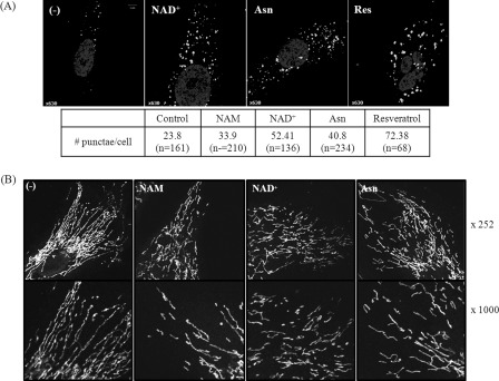
FIGURE 3.
Increase in LC3 puncta and mitochondrial fragmentation in the cells treated with NAD+ or Asn. A, cells cultured on a coverslip were either mock-treated (−) or incubated in the presence of 5 mm NAM, 5 mm NAD+, 10 mm Asn, or 10 μm resveratrol for 2 days and immunostained with antibody against LC3B protein (white) and counter-stained with Hoechst 33258 for nuclear DNA (gray). Representative confocal microscopic image of a cell is presented. The numbers of prominent LC3 puncta (bigger than 0.5 μm in diameter) in a cell were counted using the ImageJ program (rsbweb.nih.gov), and the mean numbers are presented in the table. B, cells cultured in the absence (−) or the presence of NAM, NAD+, or Asn for 2 days as above were stained with MitoTracker Red, fixed, and visualized in confocal microscopy. Magnifications: top row, ×252; bottom row, ×1000.

