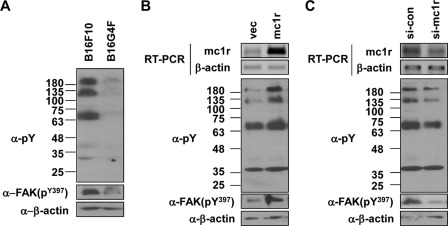
FIGURE 2.
MC1R regulates the activation of the focal adhesion kinase. A, B16G4F and B16F10 cells were distributed to tissue culture plates and incubated at 37 °C for 48 h. Total cell lysates were analyzed by Western blotting with either anti-phosphotyrosine (α-pY) or anti-phospho FAK (α-FAK(pY397)) antibodies; β-actin (α-β-actin) was detected as the loading control. B, B16G4F cells were transfected with either vector (vec) or MC1R. After 24 h, total RNA was extracted, and mRNA expressions were analyzed by RT-PCR, using β-actin as the loading control (top panel). Total cell lysates were analyzed by Western blotting as described for A (bottom panel). C, B16F10 cells were transfected with siRNA targeting MC1R (si-mc1r). After 48 h, MC1R mRNA expression was analyzed by RT-PCR. β-Actin was used as the control (top panel). Total cell lysates were analyzed by Western blotting as described for A (bottom panel). si-con, control siRNA.

