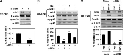
FIGURE 8.
α-MSH inhibits syndecan-2 expression via stimulation of p38 activation. A, B16F10 cells were treated with α-MSH (1 μm) for 24 h. Syndecan-2 (sdc2) mRNA expression was analyzed by RT-PCR. β-Actin was used as the loading control. The cells were lysed using radioimmune precipitation assay buffer, and total cell lysates were analyzed by Western blotting with anti-phospho p38 (α-p-p38) antibody. Anti-p38 (α-p38) antibody was detected as a control (top panel). B16F10 cells were harvested and seeded into the upper chambers of Transwell plates. After 24 h, migrated cells were stained with hematoxylin and eosin (bottom panel). **, p < 0.05 versus untreated cells. B, B16F10 cells pretreated with SB239063 (SB, 5 μm) for 30 min were incubated with α-MSH (1 μm). After 24 h, syndecan-2 expression levels (top panel), phosphorylation of p38 (middle panel), and cell migration (bottom panel) were analyzed as described for A. **, p < 0.05 versus untreated cells. C, B16F10 cells transfected with MC1R were treated with α-MSH (1 μm). After 24 h, syndecan-2 and MC1R expression levels (top panel), phosphorylation of p38 (middle panel), and cell migration (bottom panel) were analyzed as described for A. *, p < 0.01; **, p < 0.05 versus control. vec, vector.

