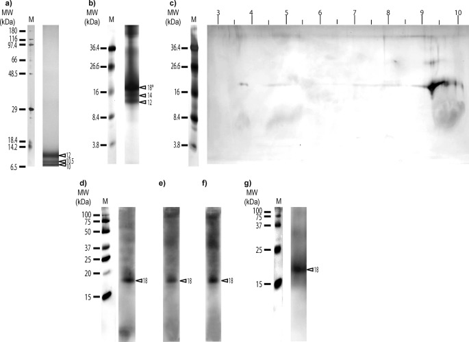
FIGURE 1.
Silver staining of electrophoresis gels and Western blots on organic matrix extracted from sclerites of C. rubrum. a, one-dimensional SDS-PAGE (10 μg protein/lane; BisTris 12% polyacrylamide gel, silver stain molecular mass marker (M6539; Sigma) as protein marker). b, one-dimensional SDS-PAGE (10 μg protein/lane, Tris-Tricine 16.5% polyacrylamide gel, kaleidoscope polypeptide standards (161-0325; Bio-Rad) as protein marker); c, two-dimensional electrophoresis gel (Tris-Tricine 16.5% polyacrylamide gel, kaleidoscope polypeptide standards (161-0325) as protein marker) of the major protein band (18*) excised from one dimensional SDS-PAGE. d, Western blot (5 μg of proteins/lane; BisTris 12% polyacrylamide gel, Precision Plus Protein WesternCTM standard (161-0376; Bio-Rad) as protein marker) with antibodies against phosphorylated serine. e, Western blot with antibodies against the phosphorylated threonine (5 μg of proteins/lane; BisTris 12% polyacrylamide gel, Precision Plus Protein WesternCTM standard (161-0376) as protein marker). f, Western blot with antibodies against phosphorylated tyrosine (5 μg of proteins/lane; BisTris 12% polyacrylamide gel, Precision Plus Protein WesternCTM standard (161-0376) as protein marker). g, Western blot with anti-scleritin antibodies (Tris-Tricine 16.5% polyacrylamide gel, Precision Plus Protein WesternCTM standard (161-0376) as protein marker). MW, molecular mass; M, protein marker; and triangles show apparent molecular mass of proteins (in kDa).

