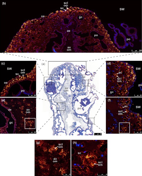
FIGURE 3.
Histology and immunohistochemistry of a longitudinal section of a demineralized branch of C. rubrum. a, overview of a section stained with hemalun/eosin/aniline blue which, respectively, stain nuclei and cytoplamic and connective regions observed with a bright field light microscope. b–h, labeling with antibodies against scleritin (orange to yellow) is merged with blue labeling of cell nuclei with DAPI: b, immunolabeling of apical region of the colony; c, immunolabeling of tissues close to a polyp; d, immunolabeling of oral epithelium; e, immunolabeling at the interface between aboral epithelium and organic matrix of axial skeleton; f, immunolabeling at the base of a branch; g, magnification of e showing labeling of organic matrix of sclerites inside the organic matrix of axial skeleton. h, magnification of f showing labeling of organic matrix of a sclerite inside a scleroblast. Observations from b to h were performed with a confocal microscope. SW, sea water; po, polyp; oe, oral epithelium; sc, scleroblast; gc, gastrodermic canal; ae, aboral epithelium; ax om, organic matrix of axial skeleton; scl om, organic matrix of sclerite. Each image was obtained by Z-stack reconstruction of 10 tissue sections, with 1-μm step.

