FIGURE 4.
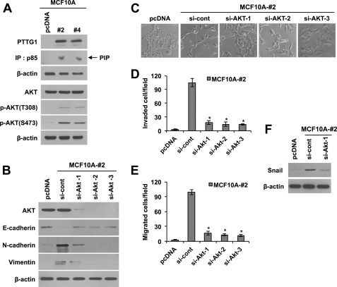
PTTG1 induces EMT via PI3K/Akt signaling. A, Western blots. PTTG1 expression led to an increase of phosphorylated AKT at Thr-308 and Ser-473 in MCF10A cells. PI3K assay showed that PTTG1 expression increases the activity of p85, a component of PI3K. β-Actin was used as the loading control. IP, immunoprecipitation. B, Western blots. Transfection of PTTG1-expressing MCF10A (clone #2) with three different Akt-targeting siRNAs decreased epithelial marker, E-cadherin, and increased mesenchymal markers, N-cadherin and vimentin. β-Actin was used as the loading control. C, transfection of PTTG1-expressing MCF10A (clone #2) with three different Akt-targeting siRNAs recovered cell morphology to MCF10A cells. Representative phase-contrast images are shown. D and E, treatment with three different sRNA targeting AKT suppressed invasive (D) and migratory properties (E) of PTTG1-expressing MCF10A cells (clone #2). Invasive and migratory properties were analyzed in trans-well by counting migrated cells in randomly selected five microscopic fields per well. Error bars represent mean ± S.D. of triplicate samples. *, p < 0.001, versus control. F, Western blots. Treatment with siRNA targeting AKT attenuated PTTG1-induced expression of Snail in PTTG1-expressing MCF10A cells (clone #2). β-Actin was used as the loading control.

