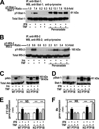
FIGURE 3.
Inhibition or absence of PTP1B restored the response to IFNα in cells treated with TNFα. A, HepG2 cells were exposed to 250 units/ml IFNα for 60 min, 20 ng/ml TNFα for 18 h, or both in the absence or presence of 0.1 mm pervanadate added to the cells 60 min before treatment with IFNα. Cellular proteins were immunoprecipitated (IP) as indicated in Fig. 1C, and tyrosine-phosphorylated Stat-1 was analyzed (pY-Stat1) by Western blot (WB). B, HepG2 cells were exposed to 100 nm insulin (Ins) for 15 min, 20 ng/ml TNFα for 18 h, or both in the absence or presence of 0.1 mm pervanadate added to the cells before insulin. Cellular proteins were immunoprecipitated with anti-IRS-2 antibody analyzed by Western blot using specific antibody against IRS-2 and antiphosphotyrosine. pY-IRS-2, IRS-2 tyrosine-phosphorylated. C, neonatal PTP-1B-deficient hepatocytes (KO PTP1B) and their counterpart wild-type cells were cultured (WT PTP1B) in the presence or absence of 20 ng/ml TNFα for 18 h and treated with 100 nm insulin for 15 min. Total and serine phosphorylated Akt was analyzed by Western blot. D, total and tyrosine-phosphorylated Stat-1 were measured in neonatal wild-type and PTP-1B-deficient hepatocytes cultured in the conditions described in panel C and treated with 250 units/ml IFNα instead of insulin. E and F, neonatal PTP-1B-deficient hepatocytes were treated as indicated in panel D. 2′5′ OAS (E) and Mx (F) gene expression were measured by quantitative RT-PCR using endogenous GAPDH as reference. ***, p < 0.001; NS, not significant.

