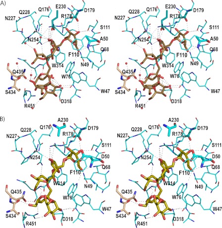
FIGURE 4.
Binding mode of the substrates. A, stereo view of the D50A active site with important protein residues engaged in inulin recognition. The major interactions are indicated by a dotted line. Inulin is colored in brown and residues from the adjacent subunit B in beige. The catalytic triad is represented by thicker sticks. Water molecules are shown as red spheres. B, stereo view of the Glu-230 active site showing the fructosylnystose in lime green. Direct polar links between the substrate and the enzyme are shown as dotted lines.

