
Figure 1. Effect of polyP on the barrier permeability of endothelial cells.
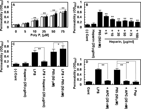
(A) Endothelial cells were incubated with indicated concentrations of polyP comprised of 45 (white bars), 65 (grey bars) and 70 (black bars) phosphate units for 4h followed by measuring permeability as described in Methods. (B) The same as panel A except that permeability was monitored with polyP65 after treating endothelial cells with indicated concentrations of unfractionated therapeutic heparin for 4h. (C) The same as above except that cells were incubated with LPS (10 ng/mL for 4h) with or without prior incubation with either heparin (20 μg/mL for 4h) or polyP65 (50 μM for 4h). (D) The same as above except that cells were pre-incubated with APC (20 nM for 3h) before treating cells with either LPS (10 ng/mL for 4h) or polyP65 (50 μM for 4h) in the absence or presence of 0.05 U/mg phosphatase (Psp). All results are means ± SD of three different experiments. *p < 0.05; **p < 0.01 compared to 0 (A), polyP65 (B), LPS (C).