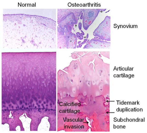
Figure 2.
Histologic features of osteoarthritis (OA). The normal synovium has a thin (1–2 cells thick) lining layer and a vascularized, loose connective tissue sublining layer. OA synovium demonstrates features of synovial villous hyperplasia (#), lining hyperplasia (arrows), increased vascularity (+) and perivascular mononuclear cell (inflammatory) infiltration. In OA articular cartilage, loss of cells and matrix is accompanied by areas of cell clusters. There is thickening of the calcified zone and duplication of the tidemark which normally separates articular cartilage from the underlying calcified cartilage. The subchondral bone is also thickened and vascular invasion, which can extend through the tidemark and into the base of the articular cartilage, is seen. Histology kindly provided by Ed DiCarlo, Hospital for Special Surgery, New York, NY.

