
Figure 1.
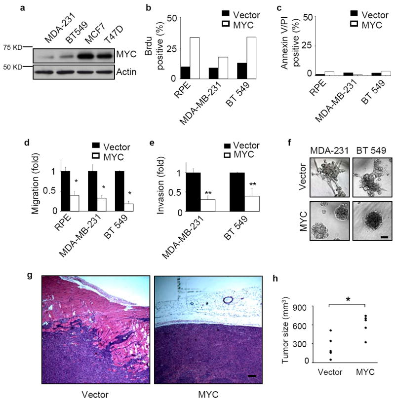
Elevated MYC expression impedes the invasiveness of human breast cancer cells. (a) MYC expression levels in four breast cancer cell lines. The endogenous MYC expression levels of MDA-MB-231, BT549, MCF7, and T47D cells were measured by western blots. (b-c) Ectopic overexpression of MYC increases proliferation but does not affect apoptosis. Cells were labeled with Brdu on tissue culture plastic for 30 minutes at 37°C (b, n=2) or incubated with annexin V and propidium iodide (PI) for 5 minutes at room temperature (c, n=2). Both positive and total nuclei were counted and the results are expressed as mean±SD, * p<0.05. (d) MYC overexpression inhibits cell migration. Migration of MDA-MB-231, BT549 and RPE cells was measured in Boyden-chamber assays (n=3 for each experiment). The cells were transfected with either vector control or a MYC construct. (e) The invasiveness of breast cancer cells is inhibited by MYC overexpression. Cell invasion through Matrigel-coated transwells was measured for MDA-MB-231 (n=3) and BT549 (n=3) cells stably transduced with vector control or exogenous MYC. (f) High level of MYC expression abrogates the invasive phenotype of breast cancer cells grown in 3D Matrigel. Results are shown for day 6. Scale bars: 50 μm. (g-h) MYC overexpression enhances tumor growth but reduces tumor invasion into nearby tissues. MDA-MB-231 cells stably expressing vector or exogenous MYC were inoculated subcutaneously into nude mice. The tumors were collected 4 weeks after injection, sectioned and stained with hematoxilin and eosin (H&E, g) and assessed for size (h, n=5 for each group, p<0.01). Scale bar: 200 μm. Results are expressed as mean± SD. * p<0.05; ** p<0.01.