
Figure 5.
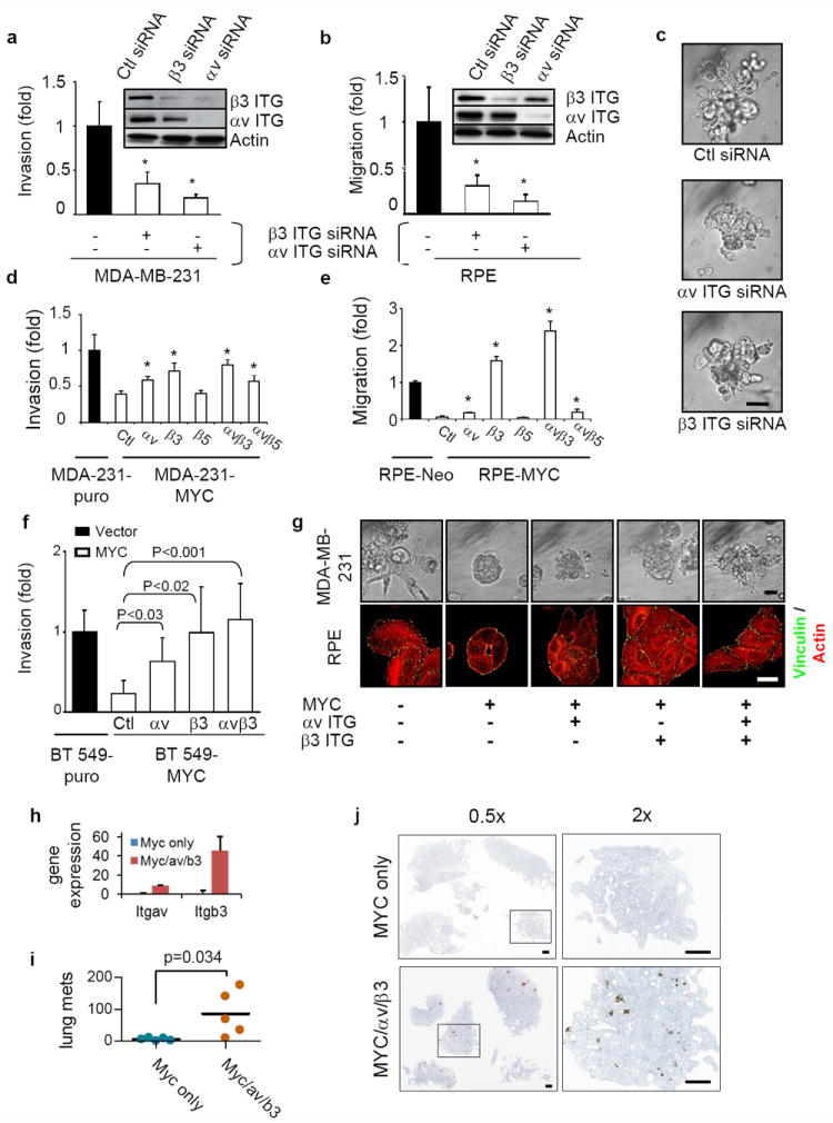
MYC affects breast cancer cell invasiveness by suppressing integrin αv and β3 subunits. (a-b) Knockdown of αv or β3 integrin inhibited invasion (a) and migration (b) in a Boyden-chamber assay, n=3. Knockdown of the integrins by siRNA was confirmed by western blots. (c) Knockdown of αv or β3 integrin inhibits the invasiveness of MDA-MB-231 cells grown in a 3D Matrigel assay for 6 days, n=3. Scale bar: 50μm. (d-e) αv and β3 integrin rescues the compromised migration (d) and invasiveness-(e) elicited by high MYC expression, n=3 for each. MDA-MB-231 and RPE cells overexpressing MYC were transiently transfected with vector, αv, β3, or β5 integrin constructs. Cell invasiveness and migration were assessed by Boyden chamber assay. (f) Inhibition of cancer cell invasiveness by MYC over-expression can be rescued by exogenous expression of αv and β3 integrin subunits in BT549 cells (n=3). (g) Expression of exogenous αv and β3 integrin partially rescued actin cytoskeleton, focal adhesion formation of RPE cells grown on 2D tissue culture plastic dishes, and the compromised invasiveness of MDA-MB-231 cells in a 3D Matrigel assay. RPE or MDA-MB-231 cells, stably expressing the indicated constructs, were plated on cell culture dishes for 24 hours or in 3D Matrigel for 6 days. RPE cells were then stained with anti-vinculin antibody (green) and Texas-Red-conjugated Phalloidin (red). Images of 3D Matrigel culture were obtained by phase contrast microscopy. Scale bars: 50 μm for phase contrast and 5 μm for immunofluorescence staining. (h) Quantitation of Itgav and Itgb3 transcripts by quantitative PCR in MDA/MYC and MDA/MYC/αv/β3 cells, n=3. (i) Quantitation of increased lung metastases in mice orthotopically implanted with MDA/MYC/αvb3 cells as compared to MDA/MYC cells, n=5 for each. (j) Images of lungs of mice orthotopically implanted with MDA/MYC/αvβ3 or MDA/MYC cells. Results are expressed as mean± SD. *, p<0.05.