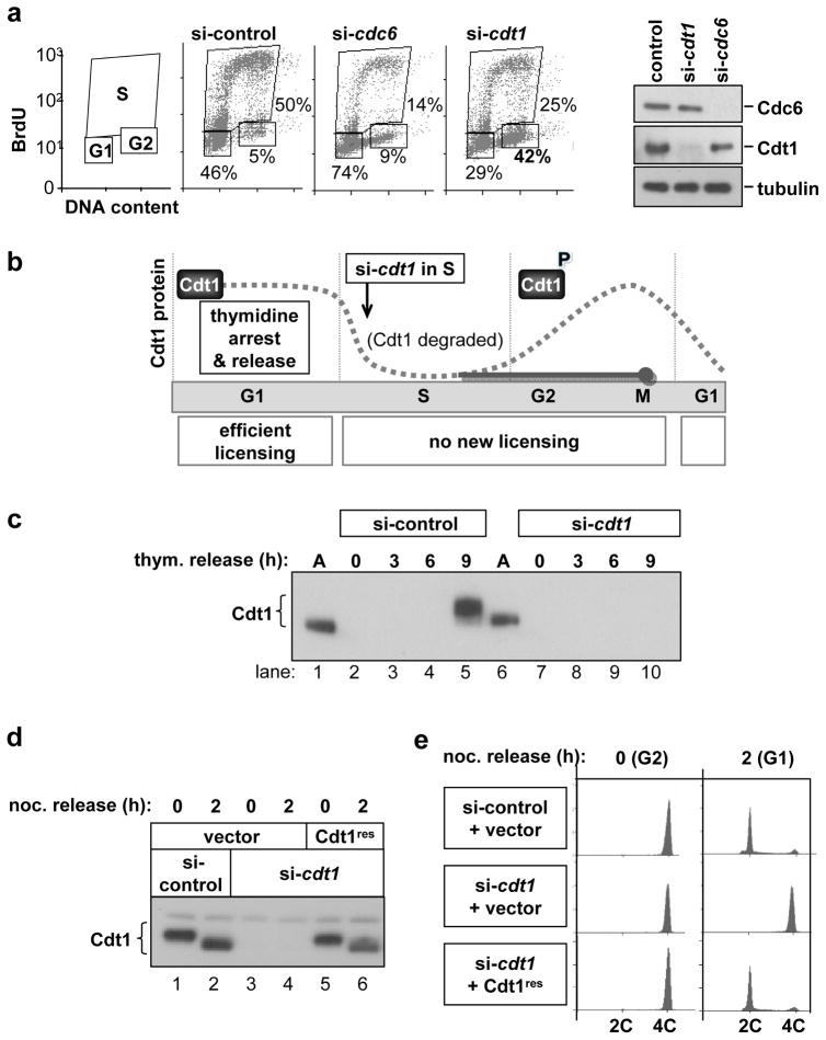
Figure 1. Cells depleted of Cdt1 after S phase do not complete cell division.
(a) Normal human fibroblasts (NHF1-htert) were transfected with siRNAs targeting cdc6, cdt1, or GFP (control) mRNAs for 72 hours. Cells were labeled with BrdU for the final hour and then analysed by flow cytometry for cell cycle position and by immunoblotting for endogenous Cdc6, Cdt1, and Tubulin. (b) Diagram of experimental design. Endogenous Cdt1 protein levels normally drop during S phase due to ubiquitin-mediated proteolysis and recover beginning in G2 (dotted gray line). Release from an early S block into cdt1 siRNA transfection medium blocks Cdt1 protein re-accumulation (solid gray line). (c) Immunoblot analysis of endogenous Cdt1 protein in cells synchronized as diagrammed in b; M phase occurs between 9 and 10 hours post-release from the second thymidine arrest. (d) Stable HeLa cell lines transduced with empty vector (lanes 1–4) or an siRNA-resistant form of Cdt1 (“Cdt1res” lanes 5 and 6) were synchronized as in b and transfected with either control siRNA targeting GFP (lanes 1 and 2) or with cdt1 siRNA (lanes 3–6). Cells were released from early S phase into nocodazole for 10 hr and then either harvested (0 hr) or released for 2 hours into G1 phase, and Cdt1 protein levels were analysed by immunoblotting. (e) Flow cytometric analysis of DNA content of cells in d.

